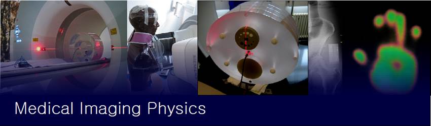
Medical imaging physics
covers different modalities such as ultra sound, magnetic resonance tomography,
diagnostic nuclear medicine (PET, SPECT), Fluoroscopy (XRF), Angiography (XRA) or
CT. Actual research activities in the group can be split into the following
fields:
Optimisation
of CT protocols: Optimisation of CT protocols is important
to ensure a sufficient image quality with a radiation exposure as low as
possible. Recently, image quality and radiation exposure (especially dose-length product DLP) are
simultaneously evaluated by the KSA group using the ZHAW elliptical phantom.
Complementary to this information, patient exposure data (CTDI, DLP) from CT
units can be collected automatically by dedicated software such as DIDT or GE DoseWatch. The calibration of the dose indications of the
CT unit can be
checked by a CTDI phantom. In consequence, DLP and CTDI are available for
patients and phantom
measurements for a large variety of clinical protocols and CT units. For the
same protocols, image quality data is only available for the phantom
measurements. To elaborate a base line for of the CT protocols concerning image
quality and dose, the collection of intrinsic image quality of patient scans is
required to complete the information needed for a quantitative basis form
optimisation. Actual research includes the development of novel phantoms and
software algorithm for evaluating low contrast detectability and clinical image
quality assessment.
Radiation
exposure of mammography and XRF: In collaboration with the Kantonsspital Baden (KSB), the radiation exposure with and
without different protective devices is evaluated by dosimetric
measurements and Monte-Carlo (MC-) simulations as well. A similar project in
collaboration with the ETH Zurich is dedicated to the investigation of the
radiation exposure during fluoroscopy of hip-implants using a XRF-robot.
Physiological
parameter assessment using MRI-based techniques:
Our group is experienced in developing dedicated phantoms for calibration and
QA of physiological measurements. This knowledge can be combined with the
experience in modelling physiological processes and model-based data analysis.
Nuclear
medicine: We focus especially on pharmacokinetic- and pharmacodynamic modelling in combination with imaging data.
Risk
assessment and radiation protection: We combine radiation
measurements with MC- simulations and dedicated dynamic models to assess
radiation risks from different sources such as X-ray units, linear accelerators
or radioactive isotopes.
References:
Özden
I, Scheidegger S (2014): The influence of scan length and collimation on the CTDIvol measurement in case of helical CT. Proc. of Joint Conference of SSRMP, DGMP and
ÖGMP 2014 (ISBN 987-3-9816508-5-3), 459-460
Scheidegger
S, Marder D, Timm O, Bonmarin M, Rhodes S (2014): Measuring skin perfusion after
superficial hyperthermia using IR-cam technology. SSBE Annual Meeting 2014, 20
Haller
K, Markert B, Lutters G, Scheidegger S (2013): Dosimetric comparison of the elliptical ZHAW-CTDI-phantom
with the standard CTDI phantom. SSRMP
annual scientific meeting 2012, P07, 58
Haller
K, Scheidegger S, Lutters G (2012): Linac radiation
shielding under clinical conditions – radiation protection case report. Proc. of SSRMP annual scientific meeting 2012,
56
Boye D, Müller B, Tobler S, Wassmer F,
Buck M, O’Brian K, Remonda L (2013): Quality
assurance phantom for MRI-based flow measurements. SSRMP annual scientific meeting 2012, O25, 40
Vieira
LDA, Markert B, Scheidegger S (2013): CTDI
measurements for clinical protocols and comparison to CTDI indications of CTs. Swiss Congress of Radiology 2013, 57
Scheidegger
S, Haller K, Markert B, Güdel
H, Vieira LDA, Lutters G (2013): Potential of optimisation of CR- and DR-units.
Swiss Congress of Radiology 2013, 57
Haller
K, Markert B, De Abreu Viera
L, Lutters G, Scheidegger S (2012): Evaluation of the RADPAD radiation
protection shielding using an Alderson phantom in a clinical situation
(cardiology). Proc. of SSRMP annual
scientific meeting 2012, 57
Fässler
C., Lanzolla I., Egli P., Scheidegger S.(2011): CR-based method for measuring the dose distribution
in an anthropomorphic phantom. Proc. of Dreiländertagung
ÖGMP, DGMP & SGSMP Wien, 96
Meola A., Lerch
C., Egli P., Lutters G., Scheidegger S.
(2011): Quality assurance of surface hyperthermia using a TLC-based
measurement system. Proc. of SSBE annual meeting 2011, 38
Scheidegger,
S. (2009): Comparison of image quality between a digital panorama X-ray unit with
a CdTe-CMOS detector and panorama X-ray units with
other types of digital detectors. Proc.
of SSRMP Annual Scientific Meeting 2009, 63-69.
Spaeth, N., Wyss, M. T., Weber, B.,
Scheidegger, S., Lutz, A., Verwey, J., Radovanovic, I., Pahnke, J.,
Wild, D., Westera,
G., Weishaupt, D., Hermann, D. M., K